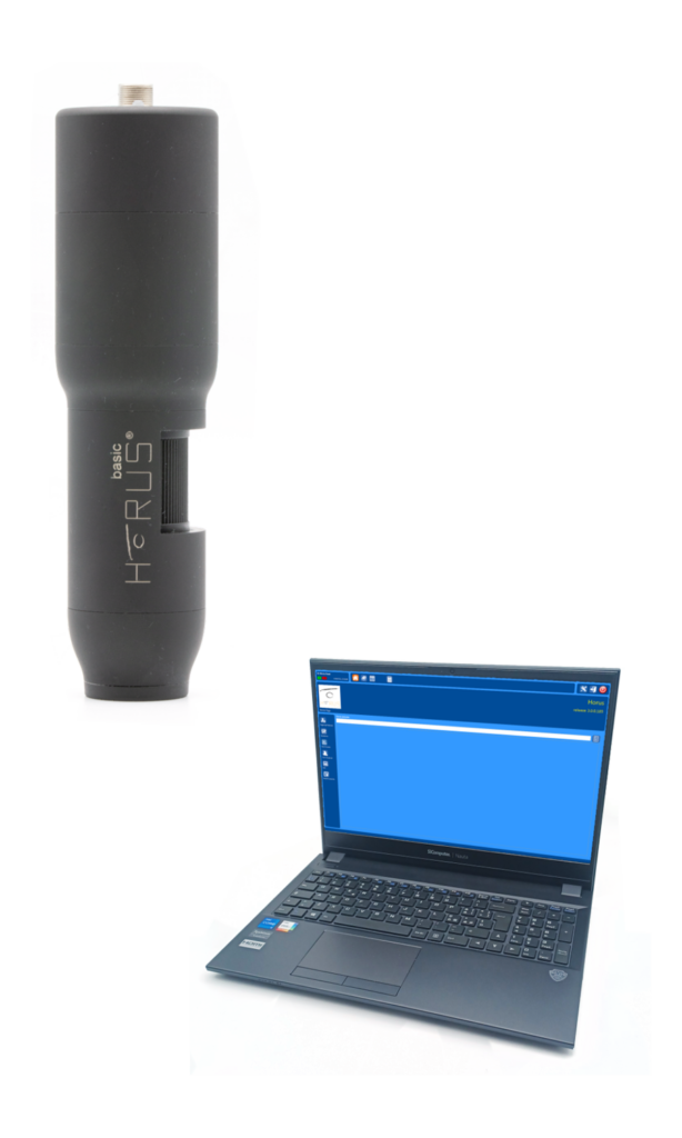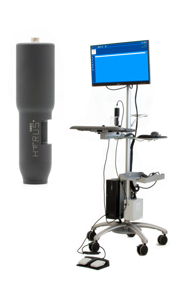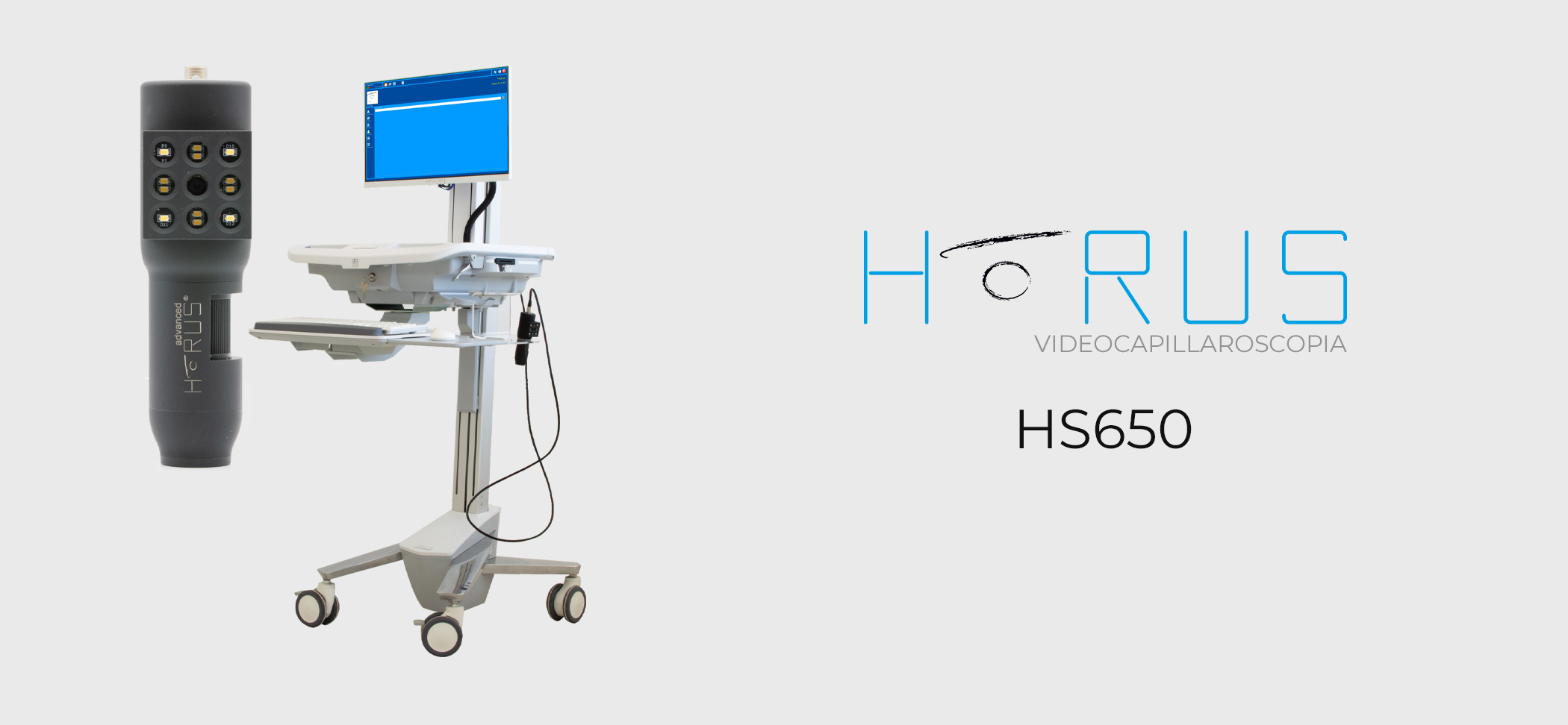
Capillaroscope Horus HS650
The first system with 4K ULTRA-HD resolution.
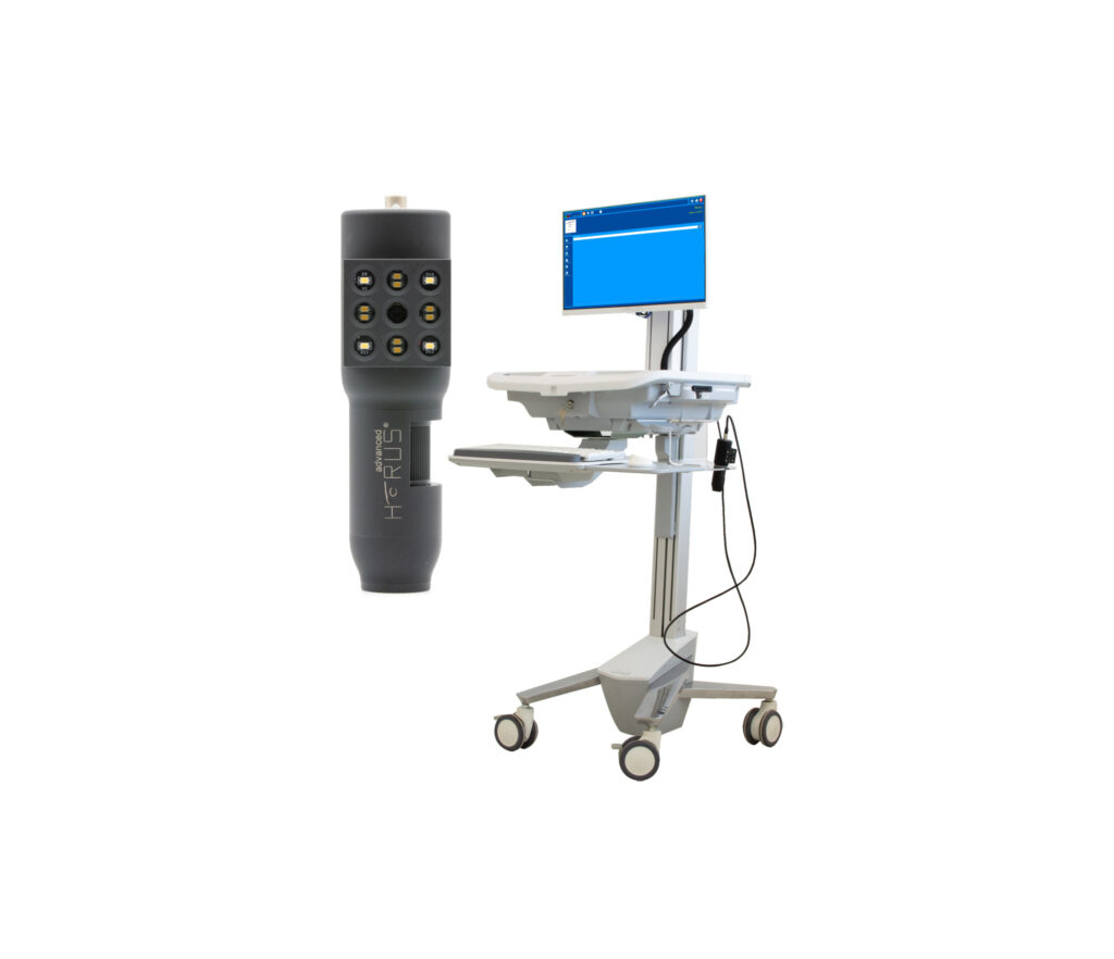
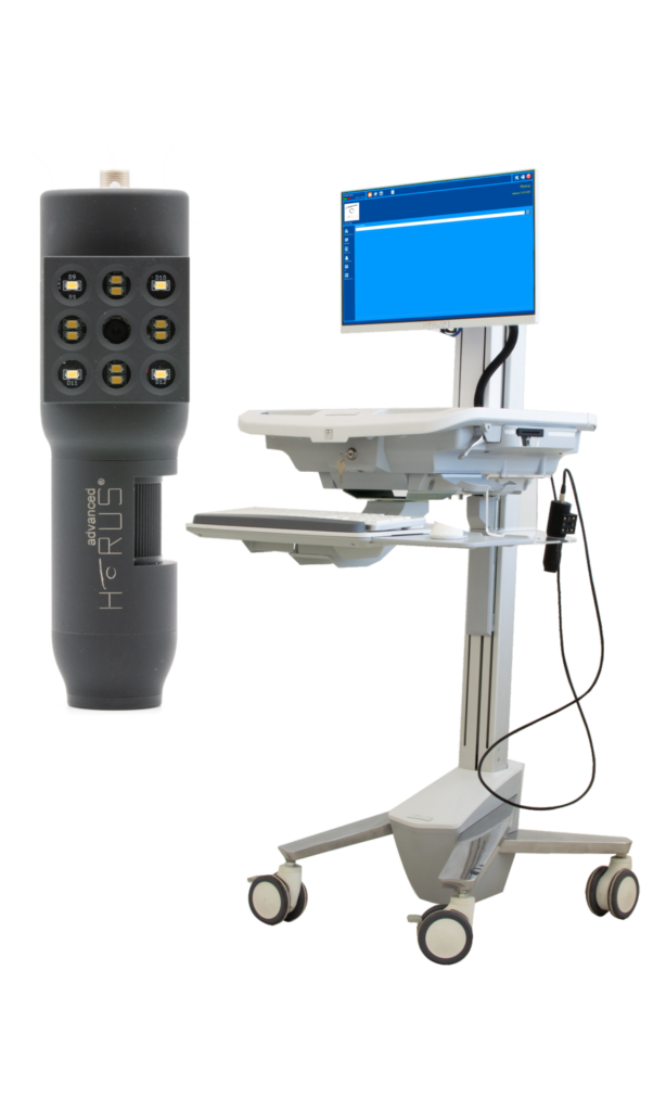
Ergonomic and compact to manage it as you wish.
It allows you to adapt the work surface, monitor, keyboard to your needs for better working condition.
Large, adjustable, built-in 22-inch monitor with Multi-finger Touch-Screen technology for quick selection of functions. Double anti-static rubberized wheel with foot pedal lock for quick and safe handling. Dedicated integrated and modular electronics with proprietary firmware to ensure maximum reliability and ease of operation.
Self-calibrating 5th generation probes for maximum reproducibility in follow-up.
Ability to connect multiple probes simultaneously with convenient probe holders to have all advanced HORUS diagnostic technologies, such as FAV, available at all times.
Integrated Wi-Fi network to connect client terminals and printers.
Possibility of WiDi (Wireless Display) Technology for wireless connection of secondary monitors.


Classical videocapillaroscopy has established itself as a key diagnostic tool for the diagnosis and clinical and therapeutic follow-up of systemic sclerosis.
Defining patterns of microvascular damage allows for precise diagnostic and/or prognostic assessments.
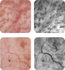
VIDEOCAPILLAROSCOPE | white light and fluorescence
The new Fluorescent Advanced Videomicroscopy (FAV) method, based on the principle of nonradiative fluorescence, allows the microcirculation to be emphasized for better morphological and dynamic assessment.
This also makes it possible to perform the examination in other anatomical districts that are easier to evaluate, highlighting capillaries otherwise invisible to classical videocapillaroscopy.
VIDEOCAPILLAROSCOPE | Measurement / Post Processing

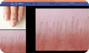
Clinical image acquisition allows detailed mapping of the areas examined making follow-up more accurate and faster.
The availability of probes with different magnifications (FOV Field Of View-from 7.5mm down to 0.340mm) allows the visualization of very accurate morphological details.
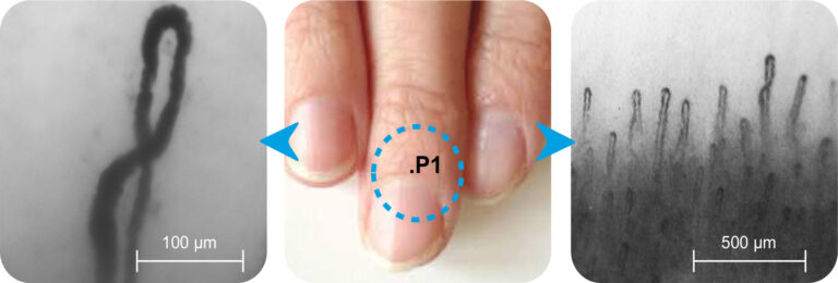
Classical videocapillaroscopy has established itself as a key diagnostic tool for the diagnosis and clinical and therapeutic follow-up of systemic sclerosis.
Defining patterns of microvascular damage allows for precise diagnostic and/or prognostic assessments.
VIDEOCAPILLAROSCOPE | white light and fluorescence
The new FAV method
(Fluorescent Advanced Videomicroscopy) based on the principle of nonradiative fluorescence, allows the microcirculation to be emphasized for better morphological and dynamic assessment.
In this way, it is also possible to perform the examination in other anatomical distres, which are easier to evaluate, highlighting capillaries otherwise invisible to classical videocapillaroscopy

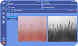
VIDEOCAPILLAROSCOPE | Measurement / Post Processing

The acquisition of clinical images permeates to effeuate a deagliated map of the examined areas making follow-up more accurate and faster.
The availability of probes with differen magnimen (FOV Field Of View- field of view- from 7.5mm up to 0.340mm) permeates the visualization of very accurate morphological deagli.


TECHNICAL DATA SHEET.
-RESOLUTION 4K ULTRA HD
-PROBE 5TH GENERATION
-INTEGRATED HIGH-BRIGHTNESS LED MODULE WITH WHITE AND POLARIZED LIGHT WITH AUTOMATIC SELECTION
-INTEGRATED MACRO AUTOFOCUS MODULE WITH WHITE AND POLARIZED LIGHT WITH AUTOMATIC SELECTION
-ASSISTED MAPPING
-AUTOMATIC FOLLOW-UP WITH REAL-TIME COMPARISON
-DIGITAL CONTROL OF ALL PROBE FUNCTIONS
-DOUBLE BLUETOOTH FOOTSWITCH (STOP/SAVE IMAGE AND VIDEO)
-SOFTWARE ADVANCED EDITION
-MONITOR 22″ CAPACITIVE TOUCHSCREEN MULTI FINGER 3-YEAR WARRANTY
OPTIONS
-WIDERM WIFI MODULE WITH CAMERA
-DOUBLE PATIENT MONITOR
– TRANSPORTER ADVANCED MODULE
-FLUORESCENCE “FAV” SOUNDS FOR LIVE VIDEOMICROSCOPE
-SUNDS WITH 50x – 100x – 200x INGRAMPLING
-POSSIBILITY OF INTERCONNECTION TO DICOM SYSTEM
